

Inside the cells of seemingly static plants are vibrant populations of motile organelles. Hundreds of mitochondria, energy providers of the cell, move about on their own individual journeys, and interact with each other as they go. They take small steps to explore their local cellular area, and use ‘highways’ in the cell (filaments made of actin protein) to travel over long distances quickly. Although this motion has been well characterised over the years, the mystery still remains: Why does the plant invest energy in moving these powerhouses around?
On the surface, plant mitochondria have an impossible task. On one hand, it is good for them to meet up. They can fuse and exchange mitochondrial DNA (mtDNA), proteins, and other chemicals, in an ongoing collaboration that is important for the plant. When this sharing is compromised, for example by mutations in the machinery responsible, plants grow less rapidly and less green, and may be sterile and experience other problems. On the other hand, it is good for mitochondria to stay apart. An even spread of mitochondria through the cell ensures an even supply of energy, limits the local buildup of damaging chemicals, and allows mitochondria to meet up with other cellular machinery. We thought that mitochondrial movement might be a way of getting the best of both worlds — allowing occasional meetups but also keeping mitochondria well spread in the cell. But to explore this idea we needed to understand both how real plant mitochondria move, and how different types of motion could help resolve this tradeoff.
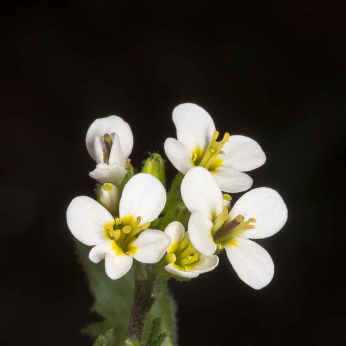
How do we delve into these communities within plant cells? Let’s start with how mitochondria move. In our lab at the University of Birmingham (our group is based at the University of Bergen but we are international!), we use live-cell laser microscopy to watch mitochondria in seedlings of Arabidopsis, a favourite plant for laboratory experiments. Prof. David Logan, who has shaped the field of plant mitochondrial dynamics, made and kindly provided us with a line of plants with fluorescent proteins attached to their mitochondria. From this, we can take videos of mitochondrial dynamics, like the time lapse below from a single cell in the hypocotyl (early stem).
A single plant cell, with mitochondria (green) moving throughout the layer of cytosol next to the cell’s surface. The outline in red is a stain showing the cell wall. A chloroplast in the centre of the cell is marked in red.
From these videos, we can use algorithms to track the positions of all the individual mitochondria over time. The computer then reports their speed, the angles at which they move, the distances between them, and the area they cover.
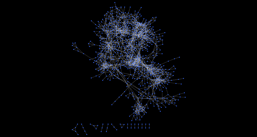
How does this movement affect their ability to meet and share contents? The answer came from a perhaps surprising perspective — social networks. Social networks describe interactions between individuals — usually people, but we applied the idea to mitochondria. When one mitochondrion comes within a small distance of another (about a micron which is a typical mitochondrion’s length), we record that ‘encounter’. These ‘encounters’ give mitochondria an opportunity to fuse and exchange contents and genetic information. Over time, these encounters build up, and can be represented as the “social” network of the population. Nodes in the network are individual mitochondria, and edges between them correspond to encounters between these organelles. Because the theory of networks like these is so well developed, we can use established theory to help us answer our questions, including: how well connected are the individuals in this cell? Do mitochondria form “cliques” (tightly-knit social groups)? How much “social” variation is there between mitochondria? And how efficient are these networks at passing information around?

We built these networks for Arabidopsis seedlings, and compared them to a wide range of computer simulations to explore what the plant could theoretically achieve with different mitochondrial motion. These simulations show that there’s indeed a tightrope to walk: a tension between mitochondria being evenly spread and highly socially connected. No plant cell, even in a simulation, can achieve both at the same time. But over time, we found that plant cells adopt a resolution to this tension that is as, or more, efficient than any of our simulations. The “efficiency” of these social networks — a measure of how easily contents can be shared between individuals — is remarkably high compared to theoretical behaviour. This suggests that the dynamics of plant mitochondria have evolved to efficiently share contents — without sacrificing their even spread through the cell, and hence their ability to deliver a uniform energy supply, avoid local build up of damaging chemicals, and meet other cellular machinery.
To support these findings, we looked at mitochondria in a mutant Arabidopsis line called friendly (so named because in this plant line the mitochondria become very “friendly”, staying together for longer times, which disturbs the even spread of mitochondria in the cell). Prof. Logan also created and provided us with plants of this line. In these plants, clustered-up mitochondria form tightly-connected cliques, which initially don’t meet other clusters very often, thus limiting their ability to share information. But interestingly, this challenge to the social-physical resolution wasn’t kept up over time. We observed that very social (popular) mitochondria travel from cluster to cluster, clique to clique, connecting these communities and eventually giving the overall network a similar efficiency to the non-mutant case.
Observing the social connectivity of these dynamic organelles has helped us uncover a trade-off the mitochondria have to deal with, and show that they use their remarkable motion to have the best of both being well-spread physically (for uniform energy delivery), and buddying up (to allow the beneficial exchange of contents) when they need to. Going forward, we are looking in more depth at the implications of this trade-off both for plant metabolism (where mitochondrial positioning shapes crosstalk with other organelles, essential for photosynthesis and photorespiration) and genetics (where mitochondrial exchange influences the maintenance and inheritance of mtDNA). These topics are of both basic biological interest and agricultural importance, as plant metabolism feeds the world, and mtDNA features play an important role in hybrid crop production.
For more information, and a more colourful look into the world of plant mitochondria, check outwww.mitochondriamove.com, and read the paper on this work here: https://www.cell.com/cell-systems/fulltext/S2405-4712(21)00133-2
Joanna and Iain are interested in the dynamics, genetics and evolution of organelles across life, and particularly in plants. The Stochastic Biology Group, which Iain leads, works with a mixture of experimental data and modelling approaches to understand complex and stochastic biological systems. Joanna is a PhD student at the University of Birmingham, UK and an avid science communicator. Iain is an Associate Professor at the University of Bergen, Norway. Follow Joanna, @ChusteckiSci, and Iain, @mitomaths, on Twitter and check out more about the Stochastic Biology Group here:https://org.uib.no/stochasticbiology/people.html. You can find more of their videos on their YouTube channel here: https://www.youtube.com/channel/UCp-q3_8CbR2Lh5PcaCYSfNQ.