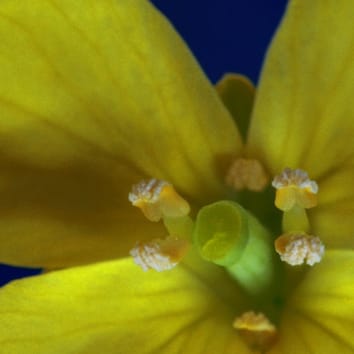


When pollen grains are released from anthers and then captured on the surface of the stigma, they obtain water and other resources from the stigma for germination and pollen tube elongation. Once the pollen tube penetrates the outer layer of the stigmatic cell wall, it grows in the apoplastic space down to the ovary for fertilization. In the ovary, two sperm cells are released from the tip of the pollen tube; one of these fertilizes the egg cell and the other the central cell, termed double fertilization, resulting in seed development. Because pollination is mediated by wind, insects and birds, pollen of other species, pathogens and dust, as well as pollen of the same species, may arrive at the stigma surface. Therefore stigmas require the ability to select suitable pollen to bring about successful fertilization.
Although pollination has been studied for many years, the molecular mechanisms involved are still largely unclear. An accurate knowledge of morphological aspects of pollination is also still far from complete. A new paper in Annals of Botany focuses on pollen behaviour during pollination. For the morphological characterization of pollination, time-lapse image analysis was used to record detailed pollen behaviour during self- and cross-pollinations in Brassica rapa. This approach demonstrated that pollen exhibits various behaviours on an individual stigma, in both self- and cross-pollinations, and the ratios of the different types of pollen behaviour are critical for successful pollination.
From these observations of pollen behaviour it is clear that supply of the correct amount of water to pollen is one of the key stages in successful pollination, and this process consists of multiple components, of hydration, rehydration and dehydration systems, and involves transport of water to and from pollen grains. The precise pollination and self-incompatibility system response in Brassica can be accomplished only when the appropriate balance and co-ordination of these processes are achieved.
Time-lapse imaging of self- and cross-pollinations in Brassica rapa. Annals of Botany (2013) 112 (1): 115-122. doi: 10.1093/aob/mct102
Pollination is an important process in the life cycle of plants and is the first step in bringing together the male and female gametophytes for plant reproduction. While pollination has been studied for many years, accurate knowledge of the morphological aspects of this process is still far from complete. This study therefore focuses on a morphological characterization of pollination, using time-series image analysis of self- and cross-pollinations in Brassica rapa. Time-lapse imaging of pollen behaviour during self- and cross-pollinations was recorded for 90 min, at 1 min intervals, using a stereoscopic microscope. Using time-series digital images of pollination, characteristic features of pollen behaviours during self- and cross-pollinations were studied. Pollen exhibited various behaviours in both self- and cross-pollinations, and these were classified into six representative patterns: germination, expansion, contraction, sudden contraction, pulsation and no change. It is noteworthy that in ‘contraction’ pollen grains shrunk within a short period of 30–50 min, and in ‘pulsation’ repeated expansion and contraction occurred with an interval of 10 min, suggesting that a dehydration system is operating in pollination. All of the six patterns were observed on an individual stigma with both self- and cross-pollinations, and the difference between self- and cross-pollinations was in the ratios of the different behaviours. With regard to water transport to and from pollen grains, this occurred in multiple steps, before, during and after hydration. Thus, pollination is regulated by a combination of multiple components of hydration, rehydration and dehydration systems. Regulated hydration of pollen is a key process for both pollination and self-incompatibility, and this is achieved by a balanced complex of hydration, dehydration and nutrient supply to pollen grains from stigmatic papilla cells.