


There are at least 4750 parasitic flowering plant species in the world, each with unique anatomy, morphology and developmental pattern. Endoparasites, which have extremely reduced vegetative bodies lacking roots, stems and leaves, make up only 1.6% of these species. They grow by embedding their cell networks in their hosts and reproduce by emerging to the outside. However, little is known about how these endoparasitic plants establish their parasitism, germinate or sexually reproduce.
Unusually, one such endoparasitic genus (Pilostyles, Apodanthaceae) seems to be missing shoot and root apical meristems entirely and has a floral meristem that develops directly from its endophytic cells. In support of these observations, Pilostyles species lack most of the genes known to be involved in shoot meristem maintenance. However, the genes involved in directing floral meristem development are present. Unfortunately, traditional studies of Pilostyles anatomy and morphology have been hampered by the species’ tiny body size.
Now, a study published in Annals of Botany by Ceccantini et al has for the first time analysed this unique genus using three-dimensional microtomography (microCT scanning) and confocal laser scanning microscopy. By analysing dozens of Pilostyles blanchetii specimens, Ceccantini et al were able to assemble a 3D visualisation of the endophytic network in its different developmental stages as well as identify the parasitic endophyte’s xylem and phloem cells.
To perform their scans, Ceccantini et al collected stems and roots of Mimosa maguirei Barneby and Mimosa foliolosa Benth. var. multipinna (Benth.) Barneby (Fabaceae) serving as hosts of the endophytic Pilostyles blanchetii (Gardner) R.Br. species as well as non-parasitised, control specimens in Serra do Cipó, Minas Gerais State, Brazil. MicroCT scanning, light microscopy, and confocal and fluorescence microscopy were performed to capture all reproductive phenophases.
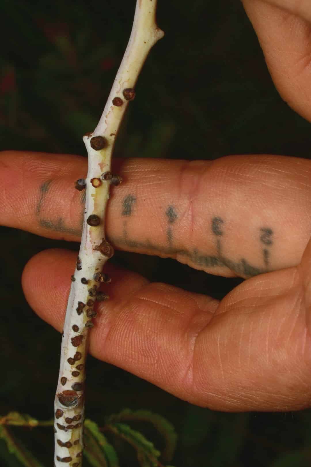
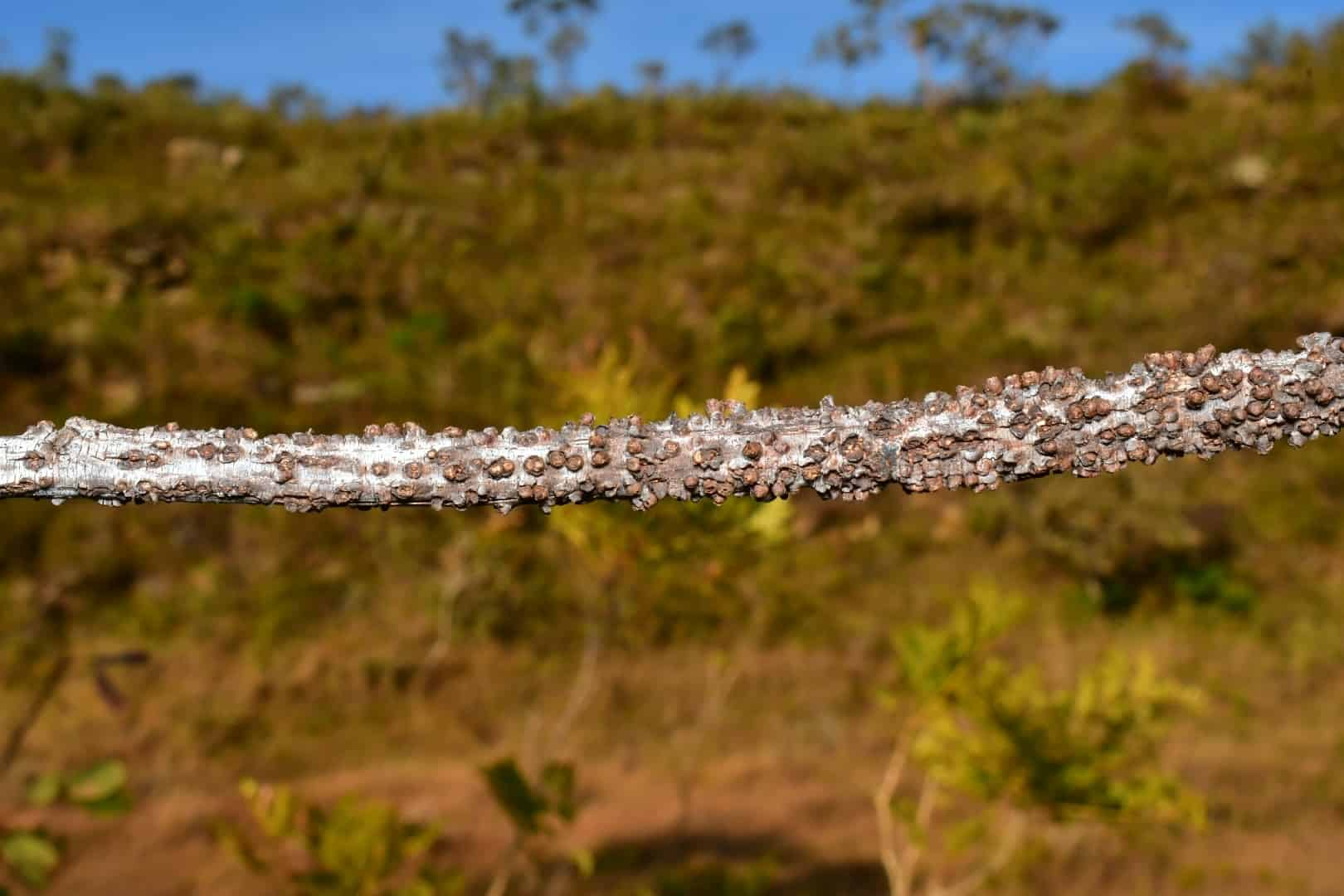
Ceccantini et al found that a host Mimosa plant is infested with “multiple ‘islands’ of parasitic tissue” derived from a single, fragmented, but inter-connected, P. blanchetii individual. Their detailed scans and microscopy also allowed the researchers to positively identify and define P. blanchetii tracheary and sieve tube elements.
“The result is a network of parasitic endophyte tissue that occupies considerable space within the host cortex and bark, which is likely to be related to the alterations in growth and development reported for parasitized Mimosa plants,” write Ceccantini et al.
Interestingly, the interconnected P. blanchetii tissues included both pistillate and staminate flowers, supporting the hypothesis that this species is monoicous. Since monoecy is rare in the Apodanthaceae family, Ceccantini et al suggest further studies combining microCT and population genetic analyses to “gain a better understanding of the evolution of sexual systems in the clade”.
Such a study would also serve to illuminate the life cycle of these rare and unusual plants.
READ THE ARTICLE
Ceccantini, G., Amaral, M.M. and Teixeira-Costa, L. (2025) “Plant life without leaves, roots, or stems: anatomy, development, and 3D structure of the endoparasite Pilostyles blanchetii (Apodanthaceae) in Mimosa hosts,” Annals of Botany, (mcaf127). Available at: https://doi.org/10.1093/aob/mcaf127
Cover image: Pilostyles blanchetii with scale in Brazil by Joey Santore / iNaturalist CC-BY-NC